Andyʼs working notes
About these notesNeuroimaging techniques
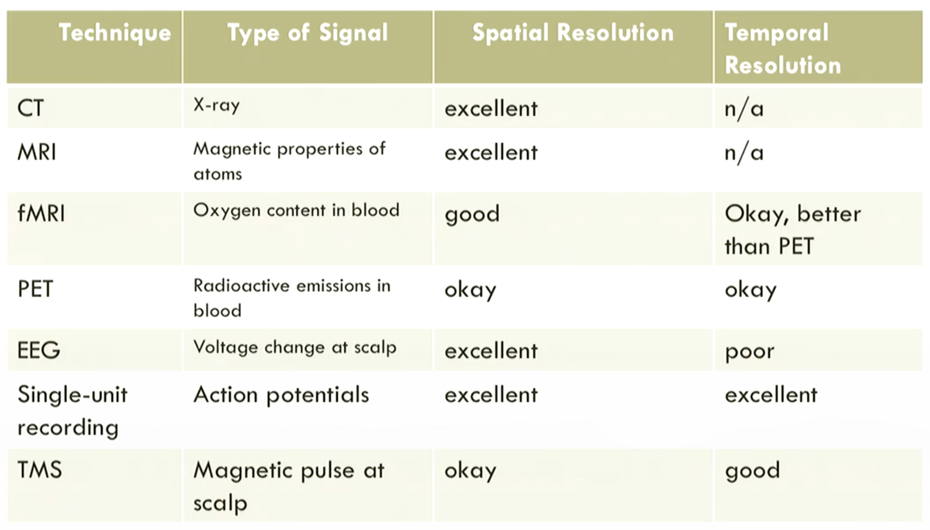
Neuroscientists choose from a number of imaging techniques according to what the situation demands.
{Structural} imaging creates pictures of {the brain’s detailed spatial layout} while {functional} imaging creates pictures of {brain activity}.
MRI and fMRI
Q. At a high level, how does MRI work?
A. By measuring magnetic interactions with the atoms that make up brain tissue.
Q. At a high level, how does fMRI work?
A. By measuring magnetic interaction with oxygen in the blood; blood flow is associated with neuronal activation.
Q. What does fMRI stand for?
A. functional magnetic resonance imaging
EEG
Q. What’s EEG stand for?
A. Electroencelphalogram.
Q. What’s the advantage of EEG over fMRI?
A. Higher temporal resolution.
Q. What’s the key advantage of fMRI over EEG?
A. Higher spatial resolution.
CT
Q. What does CT stand for?
A. computerized axial tomography
Q. Is CT for structural or functional imaging?
A. Structural.
Q. At a high level, how does CT create images?
A. By measuring how X-rays are blocked from many different angles.
PET
Q. What does PET stand for?
A. positron emission tomography
Q. Is PET for structural or functional imaging?
A. Functional.
Q. How does PET show where the brain is particularly active?
A. By introducing mildly radioactive glucose, which will concentrate in active areas.
Q. What imaging technology has largely supplanted PET imaging?
A. fMRI
Q. What’s the core trade-off between structural and functional imaging?
A. Spatial resolution vs. temporal resolution.